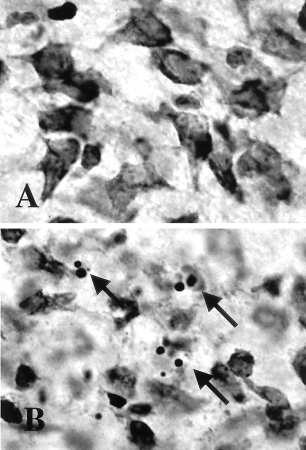
Fig. 1.

Thalamic neurons die by apoptosis after neonatal hypoxia–ischemia. A, B, Compared with sham controls (A), neurons at 24 hr after hypoxia–ischemia (B) contain many apoptotic profiles as seen in cresyl violet-stained sections of the ventral basal thalamus (arrows).
