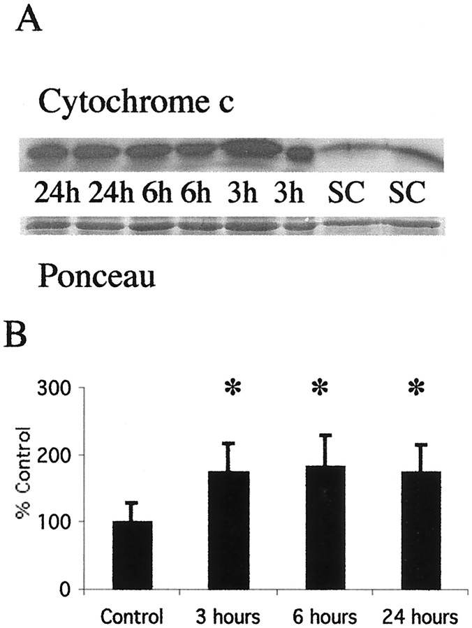
Fig. 5.
Cytochrome c accumulates in the soluble fraction in the thalamus after neonatal hypoxia–ischemia. A, Top, Immunoblot showing increased cytochrome c protein in soluble fractions from thalamic homogenates obtained 3, 6, and 24 hr after neonatal hypoxia–ischemia compared with noninjured control samples (SC). Bottom, The corresponding Ponceau-stained blot. Each lane represents a pooled sample of thalamus from three animals at the indicated time point (i.e., sham control and 3, 6, or 24 hr after hypoxia–ischemia).B, Graph showing the accumulation of cytochrome c in the thalamus over time after neonatal hypoxia–ischemia. After correcting for protein-loading differences and comparing with control, results are shown as the mean ± SD of four to five pooled samples per time point (*p < 0.05 compared with control).

