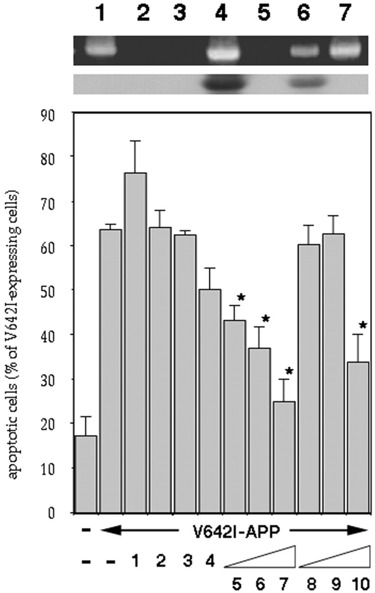
Fig. 1.

Effects of growth factors on V642I-APP-induced apoptosis. The effects of various growth factors on apoptosis in NK1 cells caused by transient expression of V642I-APP. Twenty-four hours after transfection of 0.5 μg of V642I-APP cDNA, cells were treated with or without various growth factors (lane 1, 100 ng/ml PDGF; lane 2, 10 nm EGF; lane 3, 10 nm NGF; lane 4, 10 nm IGF-II;lanes 5–7, 0.1, 1, 10 nm IGF-I; lanes 8–10; 0.1, 1, 10 nm insulin) for 24 hr, and then the percentage of apoptotic cells in V642I-APP-expressing cells was measured. Transfections were done three times independently, and the data indicate means ± SE of the three transfections. *Statistically significant versus V642I-APP transfection withp < 0.01 by Student's t test.Insets, RT-PCR clarifying the presence or absence of IGF-IR and IR in NK1 cells. In the top panel, total RNA samples (lanes 5–7), IGF-IR cDNA (lanes 3, 4), or IR cDNA (lanes 1, 2) were amplified by RT-PCR with IGF-IR primers (lanes 2, 4, 6) or IR primers (lanes 1, 3, 5), as described in Materials and Methods. Lane 7 indicates the amplified band for GAPDH as a marker of 450 bp. All amplified bands were with sizes corresponding to the expected sizes, which were 490 bp for IR and 448 bp for IGF-IR. In the bottom panel, the bands in the top panel were transferred onto nylon membrane and hybridized with IGF-IR cDNA probe.
