
Fig. 3.
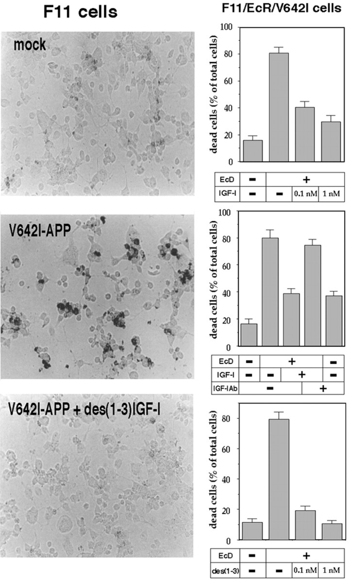
Effect of IGF-I and des(1-3)IGF-I on V642I-APP-induced neurotoxicity. In the left panels, F11 neuronal cells were transfected with or without V642I-APP cDNA and cultured in the presence or absence of 10 nm des(1-3)IGF-I. Twenty-four hours after the onset of treatment, intracellular fragmented DNA was stained in situ by TUNEL. The results indicate the representative fields. Similar experiments were repeated three times. In the right panels, F11/EcR/V642I cells were treated with or without 40 μm EcD for 48 hr in the presence or absence of IGF-I or des(1-3)IGF-I. Cell mortality was measured by Trypan blue exclusion assay. In the experiments shown in the right middle panel, cells were cultured with or without EcD in the presence or absence of 1 nm IGF-I or in the presence or absence of 5 μg/ml anti-IGF-I antibody. Values indicate means ± SD of six independent treatments.