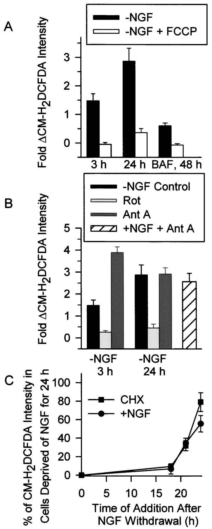
Fig. 2.

Increased ROS after NGF deprivation derived from the mitochondrial electron transport chain. A, Block of all components of the ROS burst by the protonophore FCCP (5 μm) suggested that the ROS derived from the mitochondrial electron transport chain. Cells deprived of NGF for 3, 24, or 48 hr (with 30 μm BAF) were exposed to FCCP during the time of CM-H2 DCFDA loading. FCCP was also included in the recording medium. Data were normalized as in Figure 1.n = 59–162 neurons from three to five separate platings. B, Acute effects of rotenone (Rot; 10 μm) and antimycin A (Ant A; 1 μm) on the ROS bursts at 3 and 24 hr after NGF deprivation. Also shown is the effect of Ant A on ROS in NGF-maintained neurons. Compounds were added to the culture medium during CM-H2 DCFDA loading and were also included in the recording medium. n = 59–145 neurons from three platings. C, Time course of suppression of the late ROS burst by CHX (1 μg/ml) and NGF (+NGF indicates 50 ng/ml NGF here and throughout the manuscript). Either CHX or NGF were added to the culture medium at the indicated times after NGF deprivation. The CM-H2 DCFDA intensity was then measured at 24 hr after the withdrawal. For the 24 hr time point, CHX or NGF were added during the CM-H2 DCFDA loading. Data are normalized to average CM-H2 DCFDA intensity of cells deprived of NGF for 24 hr. n = 87–163 neurons from three platings.
