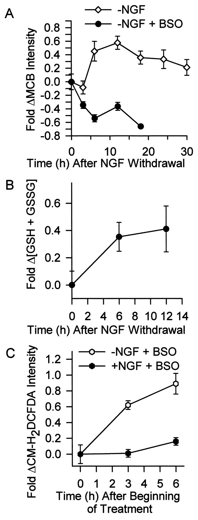
Fig. 3.

Increased GSH concentration at least partially caused the transience of the early ROS burst. A,Glutathione levels increased within several hours after NGF deprivation. The inhibitor of GSH production, BSO (200 μm), blocked this increase. Neurons were deprived of NGF for the indicated times. During the last 30 min of the incubation, cultures were exposed to the GSH-sensitive dye MCB (5 μm). Neurons were then viewed with an inverted microscope, and images were captured by a CCD camera. Intensity of MCB staining in single cells was determined with MetaMorph software and is shown as fold change from that of control cells maintained in the presence of NGF. The BSO-treated neurons appeared phase-bright and healthy for the first 12 hr after NGF deprivation. However, by 18 hr, many had died. Data are normalized to MCB intensity of NGF-maintained neurons stained and measured at the same time as the experimental cells. n = 67–137 neurons from three or four separate platings. B, The concentration of GSH plus GSSG in whole cultures increased after NGF deprivation, independently confirming the single-cell results in A. The GSH plus GSSG concentration is shown as fold increase above that measured in NGF-maintained cells sampled at the initial time point.n = 10–14 cultures from three separate platings.C, Inhibition of the GSH increase by BSO (200 μm) at least partially blocked the transience of the ROS burst after NGF deprivation. Data are shown as fold change of CM-H2 DCFDA intensity in NGF-replete or -deprived neurons exposed to BSO for the indicated times after withdrawal.n = 99–140 neurons from three or four separate platings.
