
Fig. 7.
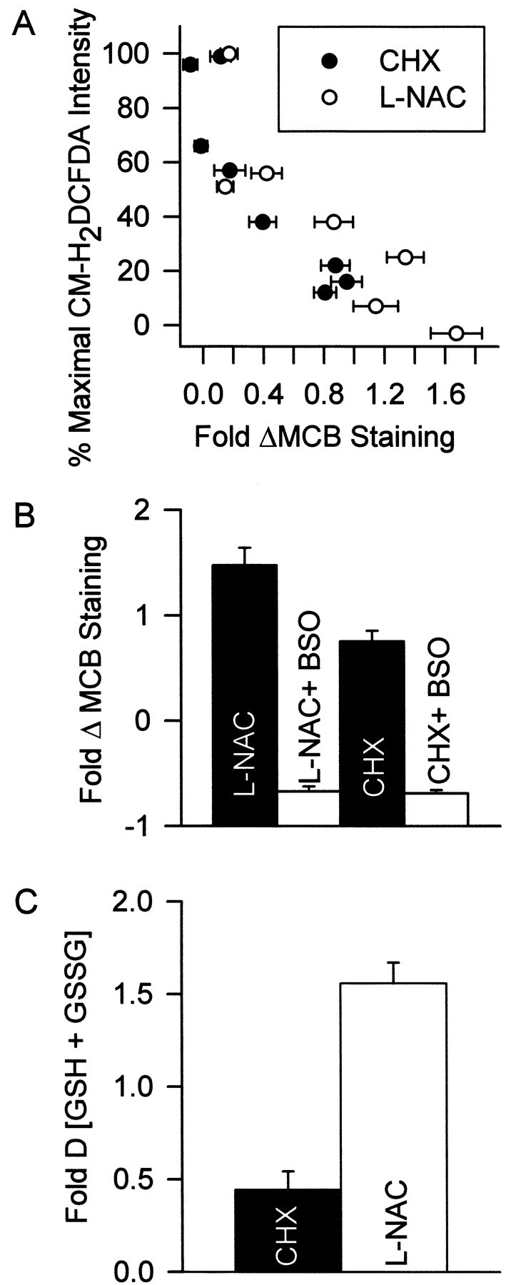
The ability of CHX and l-NAC to block the ROS burst derived from their ability to regulate cellular GSH levels. A, The abilities of different concentrations of CHX and l-NAC to block the ROS burst (CM-H2DCFDA intensity) was closely correlated with the ability of these concentrations to increase MCB staining. Cultures were deprived of NGF for 24 hr in medium containing different CHX or l-NAC concentrations and BAF (30 μm). The CM-H2DCFDA data are from Figure 6, A andB, and are matched to MCB staining resulting from treatment with the same CHX or l-NAC concentrations.n = 88–173 neurons from three separate platings.B, Increased MCB staining was caused by increased GSH concentration. The inhibitor of GSH production, BSO (200 μm), prevented CHX (1 μg/ml) and l-NAC (30 mm) from increasing MCB intensity. Cultures were treated with CHX (18 hr) or l-NAC (24 hr) before MCB staining.n = 77–123 from three separate platings.C, Effects of CHX (1 μg/ml) and l-NAC (30 mm) on GSH plus GSSG concentration in whole cultures. Cells were exposed to the compounds in the absence of NGF and BAF for 12 hr. This early time point was chosen to avoid complications from loss of cells caused by the apoptosis that would occur in the absence of BAF (Deckwerth and Johnson, 1993). Data are normalized to GSH plus GSSG concentration in cultures deprived of NGF without the compounds for the same period. n = 9–11 cultures from three separate platings for each treatment.