
Fig. 3.
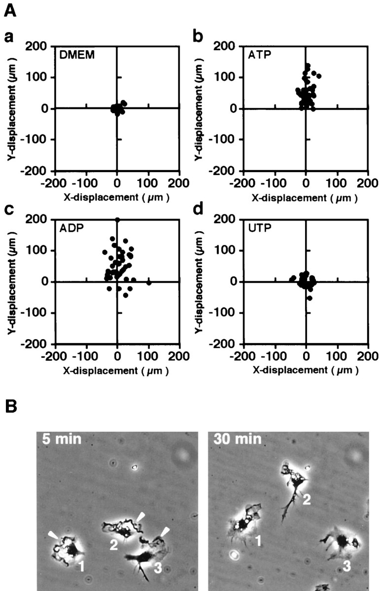
Nucleotide-induced chemotaxis of microglia in the Dunn chemotaxis chamber. A, Vector diagrams of cell displacement at 60 min after setting up the chamber. The cells were incubated in the absence of nucleotides (a) or in the presence of 50 μm ATP (b), ADP (c), or UTP (d) in the outer well. The position of the outer well of the chamber is vertically upward. All diagrams were obtained from a representative experiment using the same lot of microglial culture. We confirmed that the three independent experiments showed the same tendency. B,Displacement and morphological change at 5 and 30 min after setting up the chamber. The cells were incubated in the presence of 50 μm ATP in the outer well. The images of the same area are presented such that the position of the outer well of the chamber is vertically upward. Arrowheads indicate membrane ruffles.