
Fig. 5.
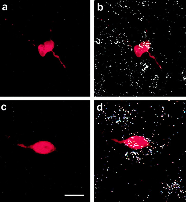
GABAergic POA neurons are identified using combined histofluorescence and in situ hybridization for GAD65 after electrophysiological recording.a, Photomicrograph of the biocytin-streptavidin-Texas Red fluorescent labeling of a recorded cell from a vehicle-treated animal. b, An overlay of the fluorescent labeling ina and the hybridization signal that clearly illustrates the double labeling for GAD65. c, Photomicrograph of the biocytin-streptavidin-Texas Red fluorescent labeling of a recorded POA neuron from an EB-treated (25 μg; 24 hr) animal. d, An overlay of the fluorescent labeling inc and the hybridization signal illustrating the double labeling for GAD65. Scale bar, ∼15 μm (for all photomicrographs).