
Fig. 1.
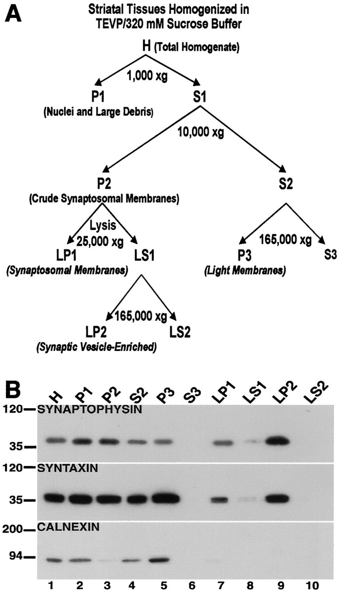
Characterization of fractionated subcellular compartments of the striatum. A, Schematic for the biochemical fractionation. The procedure for the subcellular separation of proteins as depicted in this schematic is described in Materials and Methods. B, Characterization of subcellular compartments. The isolated biochemical fractions from striatal tissues were separated by SDS-PAGE, and the blots were probed with antibodies against synaptophysin (top), syntaxin (middle), and calnexin (bottom). Synaptophysin is highly concentrated in the synaptic vesicle-enriched fraction (LP2, lane 9); syntaxin is enriched in the light membrane (P3, lane 5) and synaptic vesicle-enriched (LP2,lane 9) fractions compared with the synaptosomal membrane (LP1, lane 7); and calnexin is found in the light membrane fraction (P3,lane 5), but not in the synaptosomal membrane (LP1, lane 7) nor synaptic vesicle-enriched (LP2, lane 9) fractions.