Fig. 4.

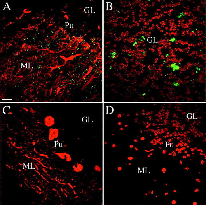
Specificity of the phosphorabphilin antibodies in immunostaining. In mice lacking Rab3A the overall level of rabphilin is reduced to ∼40% of wild type and the protein is not targeted to the synapses. We assessed the specificity of αS234-P and αS274-P by comparing their immunoreactivity in staining of brain sections obtained from wild-type and Rab3A knock-out animals. All of the panels are single confocal images taken from sagittal sections of the cerebellar cortex of adult wild-type or Rab3A knock-out mice. The staining for both phosphorylated forms of rabphilin was equivalent.A,B, Staining of sections obtained from wild-type animals; C, D, staining in Rab3A knock-out sections. The panels represent merged images of double labeling for rabphilin S234-P (green) with an antibody against calbindin (red in A and C, stains Purkinje cells) or the nuclear marker Toto-3 (redin B and D). In the cerebellar cortex of wild-type animals, phosphorabphilin was detected as a punctate synaptic-like staining in the molecular layer (A) and staining of structures in the granular layer (B). Equivalent double labeling in Rab3A knock-out sections (C, D) failed to reveal staining for phosphorabphilin, confirming the specificity of the antibodies.GL, Granular layer; ML, molecular layer;Pu, Purkinje cells. Scale bars: A–L, 20 μm.
