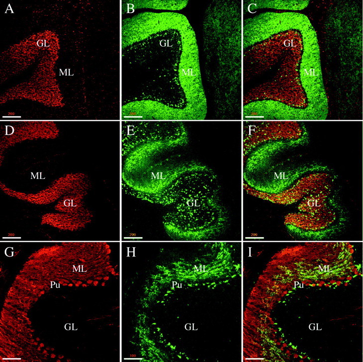
Fig. 6.

Phosphorabphilin is present in the rat cerebellar cortex. All of the panels are single confocal images taken from sagittal sections of the cerebellar cortex of adult rats. The staining for both phosphorylated forms of rabphilin was equivalent.A–C, Double staining with Toto-3 (A, stains the nuclei) and the αtotal rabphilin antibody (B, C is the merged image) shows that total rabphilin is homogeneously expressed in the molecular layer and in some structures, likely glomeruli and pinceaux, in the granular layer.D–F, Double labeling with Toto-3 (D) and αS234-P (E, F is the merged image) results in a similar staining of structures in the granular layer but a more restricted staining in the molecular layer for phosphorabphilin compared with total rabphilin.G–I, The nonhomogeneous presence of phosphorabphilin in the molecular layer is further confirmed in a co-staining for calbindin (G, stains the Purkinje cells) and rabphilin S234-P (H, I is the merged image). The staining appears punctate and concentrated in the portion of the molecular layer closest to the Purkinje cell layer. Scale bars: A–F, 200 μm; G–I, 100 μm.
