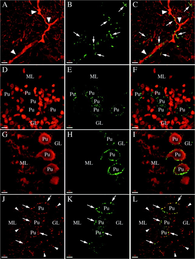
Fig. 8.

Phosphorabphilin is enriched in climbing fiber synapses. All of the panels are single confocal images taken from sagittal sections of the cerebellar cortex of adult rats (A–C) or postnatal day 8 animals (D–L). The staining for both phosphorylated forms of rabphilin was equivalent. A–C, Double staining for calbindin (A, stains the Purkinje cell dendrites) and rabphilin S234-P (B) in the molecular layer. The merged image (C) reveals that the phosphorabphilin-containing synapses (arrows) decorate the proximal aspect of the Purkinje cell dendrites (arrowheads). This staining is characteristic for climbing fiber synapses. To confirm the identity of these synapses, we double-stained brain sections of postnatal day 8 animals. At this age the climbing fibers make synapses exclusively on the cell body of the Purkinje cells, later in development these synapses disappear and new synapses are formed climbing up the Purkinje cell dendrites.D–F, Double staining with the nuclear marker Toto-3 (D, note the very large nuclei of the Purkinje cells) and with the αS274-P antibody (E). As shown in the merged image (F), phosphorabphilin staining is restricted to the cell body of the Purkinje cells.G–I, The double staining for calbindin to stain Purkinje cells (G) and for rabphilin S274-P (H) confirms the presence of phosphorabphilin-containing synapses on the cell bodies but not on the growing dendrites of the Purkinje cells (I). J–L, The synaptic nature of the staining for phosphorylated rabphilin is further corroborated by a double labeling with antibodies for the synaptic vesicle protein synaptophysin (J) and for rabphilin S274-P (K, L is the merged image). The phosphorabphilin immunoreactivity on the cell bodies of Purkinje cells colocalizes with the synaptic staining of synaptophysin (arrows), but other synapses in the molecular and granular layer (some of which are indicated by thearrowheads) are not stained for phosphorabphilin. Scale bars: A–L, 10 μm.
