
Fig. 10.
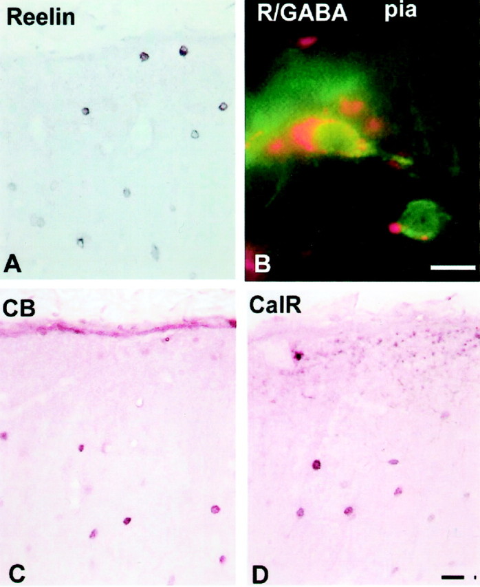
Immunohistochemistry of 5-year-old monkey visual cortex. A, Reelin-labeled cells both close to the pia and deeper in layer I. B, Double-labeling immunofluorescence with Reelin (secondary antibody conjugated with rhodamine; red) and GABA (secondary antibody conjugated with flurescine; green) revealing a C–R-like cell under the pia. C, D, Calbindin (CB)-labeled (C) and calretinin (CalR)-labeled (D) neurons in layer I. Scale bars, 20 μm.