Fig. 5.
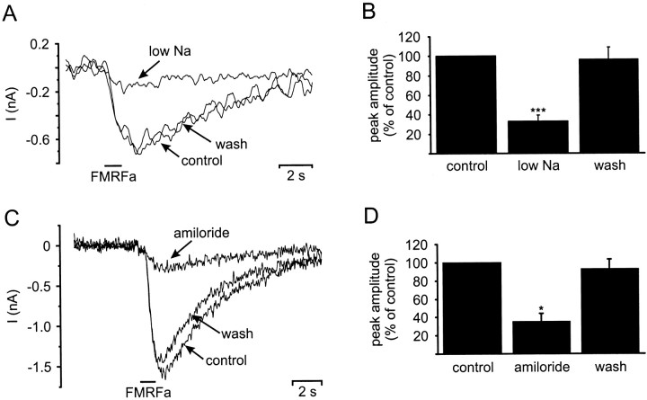
Sodium dependence and amiloride block of FMRFamide induced inward currents in isolated RPeD1 neurons. A, Replacing 90% of the extracellular Na+concentration with N-methyl-d-glucamine resulted in the reduction of the peak FMRFamide-induced (pipette concentration, 0.1 mm) inward current recorded under TEVC (holding potential, −100 mV) from −0.7 nA (control) to −0.17 nA (low Na). Returning the extracellular Na+ concentration to 50 mm (wash) reversed the effect. B, Summary of six sodium replacement experiments conducted on four cells. Reducing the extracellular Na+ concentration significantly reduced the mean peak amplitude of the FMRFamide-induced inward current to 33 ± 6% of the control value (pairedt test: p < 0.001). The mean peak amplitude returned to 96 ± 12% of the control value, when the extracellular Na+ concentration was raised again to 50 mm. C, Application of amiloride (0.1 mm) decreased the FMRFamide-induced (pipette concentration, 0.1 mm) inward current recorded under TEVC (holding potential, −100 mV) from −1.6 nA (control) to −0.3 nA (amiloride). The block was almost completely reversed after wash-out of amiloride from the bath. D, Summary of three amiloride blocking experiments. Amiloride application blocked a significant proportion of the FMRFamide-induced inward current, reducing the mean peak amplitude to 35 ± 9% of the control value (paired t test;p < 0.05). The amiloride block was reversible, and the mean peak amplitude returned to 92 ± 10% after wash-out of amiloride from the bath. *p ≤ 0.05; ***p ≤ 0.001.

