
Fig. 2.
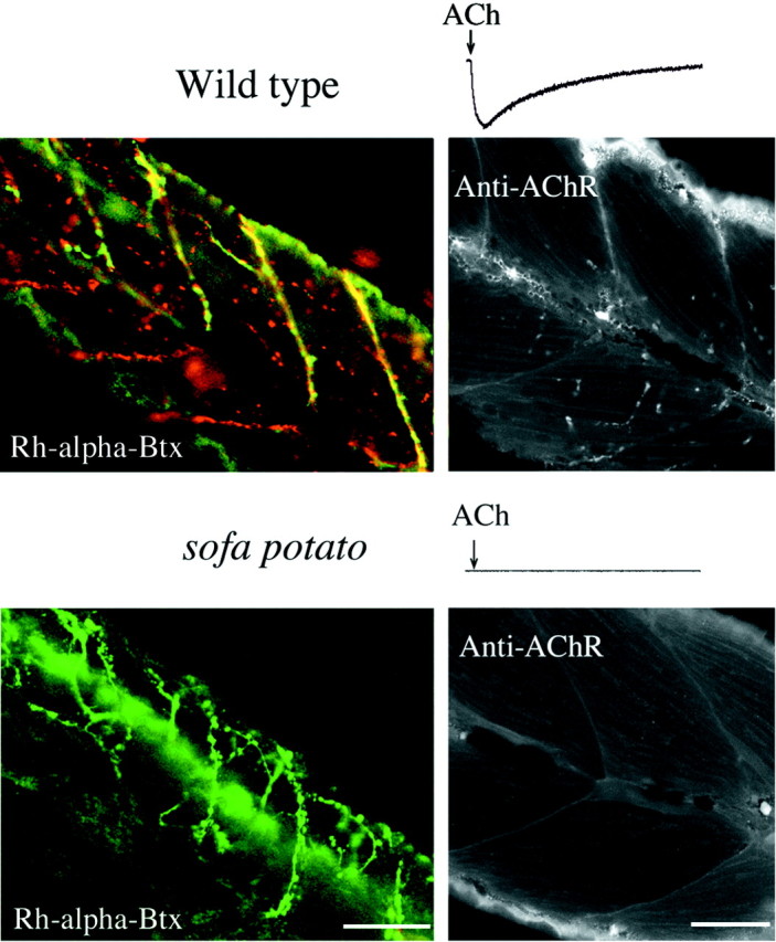
Labeling of ACh receptors by α-bungarotoxin and immunohistochemistry. Shown is the response of muscle cells from 5-d-old wild-type (top trace) and sofa potato (bottom trace) fish to transiently applied 10 μm ACh. Note the lack of response bysofa potato muscle. Muscle from wild-type/Isl1-GFP (top) and sofa potato/Isl1-GFP (bottom) lines was treated with rhodamine-α-bungarotoxin. In the top panel thered fluorescence in wild-type muscle corresponds to the location of ACh receptors. The green fluorescence from the cytoplasmic GFP indicates the location of axons and synaptic terminals. The plane of focus was set at a different level from the spinal cord so that the somata of motor neurons are not seen. Insofa potato fish (bottom) thegreen fluorescence resulting from GFP is observed, but no red fluorescence corresponding to rhodamine-α-Btx is observed. The right panels show black and white images of mAb35 labeling of ACh receptors in fixed muscle from 5-d-old fish. Some autofluorescence from the skin is seen along the edge of the tail. Fluorescence that is associated with the labeling of ACh receptors is absent in sofa potato muscle. Scale bars, 50 μm.