
Fig. 4.
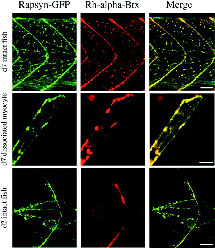
Developmental changes in the relationship between ACh receptor and rapsyn–GFP distribution. Left, Rapsyn–GFP distribution in the tail muscle of an intact 7-d-old wild-type/rapsyn–GFP fish (top), dissociated wild-type/rapsyn–GFP myocyte (middle), and 2-d-old wild-type/rapsyn–GFP fish (bottom). Thegreen fluorescence indicates the distribution of rapsyn–GFP. Middle, The distribution of fluorescence associated with the labeling of ACh receptors by rhodamine-α-Btx in the same muscle shown at the left. Right, The green fluorescence associated with the rapsyn–GFP fusion protein and the red fluorescence from the rhodamine-α-Btx-labeled ACh receptors are merged for theleft and middle images. Scale bars:Top, 50 μm; middle, 20 μm;bottom, 10 μm.