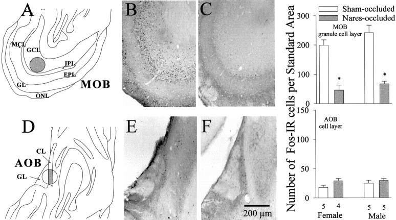
Fig. 6.
Effect of bilateral naris occlusion on neuronal Fos immunoreactivity in the granule cell layer (GCL) of the MOB and in the cell layer (CL) of the AOB of female and male ferrets killed at the end of this study after mating with an opposite-sex conspecific. The locations of the counting regions (shaded areas) are shown for the MOB (A) and AOB (D). Photomicrographs of representative examples of Fos-IR neurons in these two brain regions are shown for a sham-occluded male (B, E) and a naris-occluded male (C, F).EPL, External plexiform layer; GL, glomerular layer; IPL, internal plexiform layer;MCL, mitral cell layer; ONL, olfactory nerve layer. Right graphs, Quantitative Fos data for the MOB and AOB. The number of subjects in each group is given beneath theAOBbars. *p < 0.01,post hoc Student–Newman–Keuls comparisons with sham-occluded values of the same sex. Data are expressed as mean ± SEM.

