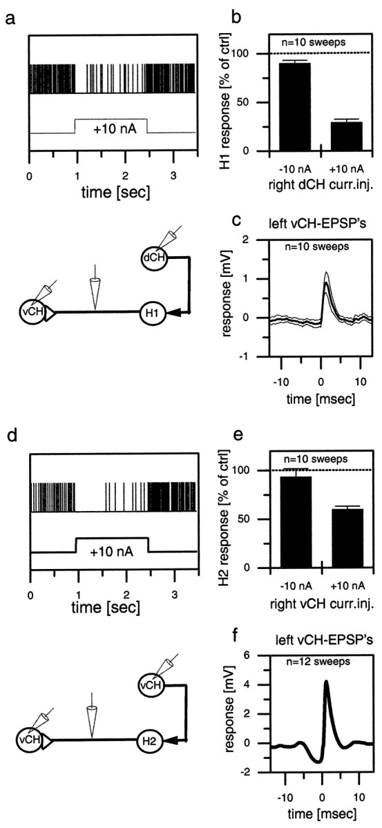
Fig. 3.

Extracellular recording of a spiking neuron and simultaneous intracellular recording of a CH cell in the same brain hemisphere (a, b, d, e) or in the opposite hemisphere (c, f). The top graphs (a–c) show results obtained from a dCH→H1→vCH cell recording; the bottom graphs (d–f) show the results for the vCH→H2→vCH cell recording. a, d, Single-responsetrace of H1 (a) and H2 (d) in response to current injected into the ipsilateral CH cell. b, e, Spike frequency of H1 (b) and H2 (e) as a function of depolarization or hyperpolarization of the CH cell. The error bars show the average and the SEM of 10 sweeps for H1 (b) and 10 sweeps for H2 (e). c, f, Spike-triggered average and SEM of the membrane potential of the contralateral CH cell, recorded within the same fly in the opposite brain hemisphere. Similar data have been obtained in four other experiments comprising all combinations at least once. They demonstrate that depolarization of the dCH as well as the vCH cell reduces spike activity in the H1 as well as the H2 cell. ctrl, Control; curr.inj., current injection.
