Fig. 4.
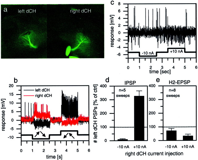
Simultaneous intracellular recordings of two dCH cells, one in the left and one in the right lobula plate.a, Fluorescence image of the cells filled with Ca-green.b, Visual responses of both cells to rotatory motion stimuli in front of the fly. c, Single-response trace of the left dCH cell in response to hyperpolarization and depolarization of the right dCH cell. Hyperpolarization led to a decreased IPSP frequency compared with resting; depolarization led to an increased IPSP frequency and a decreased EPSP frequency. d, e, These effects quantified further. Hyperpolarization of the right dCH cell suppressed IPSP frequency of the left dCH almost completely (d) but had no significant effect on its EPSP frequency (e). Depolarization of the right dCH cell led to a threefold increase of IPSP frequency in the left dCH cell (d) and decreased the EPSP frequency to ∼30% of the control condition (e).

