
Fig. 6.
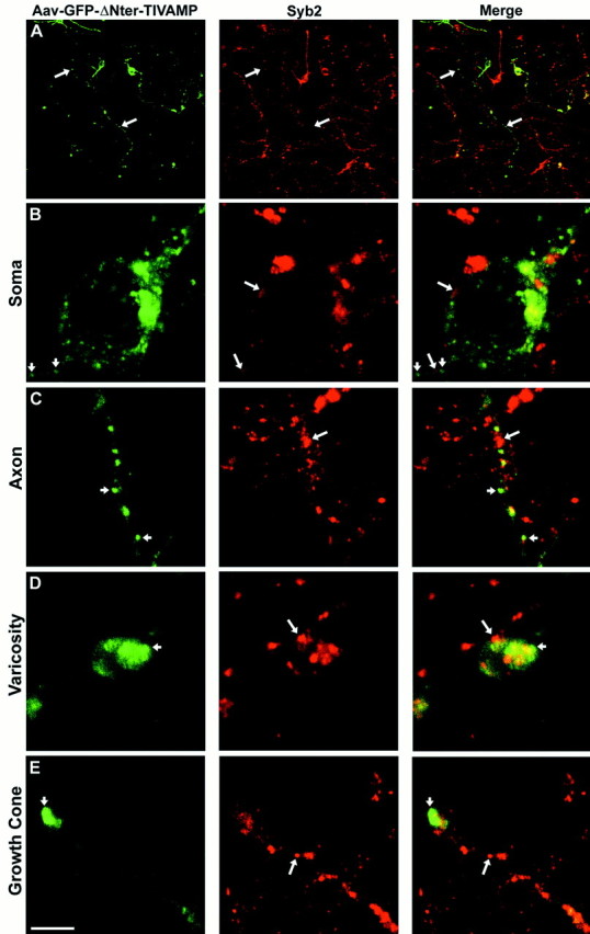
GFP-ΔNter-TI-VAMP does not colocalize with synaptobrevin 2. Rat embryonic neurons were infected with Aav carrying GFP-ΔNter-TI-VAMP. After 6 div, the cells were fixed and permeabilized, incubated with a polyclonal antibody anti-GFP and with a monoclonal antibody anti-synaptobrevin 2 (Syb2), and observed by confocal microscopy. Low magnification images are shown inA. In all the other panels high magnification images of a cell body (B), an axon (C), a varicosity (D), and a growth cone (E), respectively, are shown. GFP-ΔNter-TI-VAMP (small arrows) does not colocalize with endogenous synaptobrevin 2 (B–E, large arrows) in any of the different neuronal domains. A significant amount of GFP-ΔNter-TI-VAMP was detected at the leading edge of the growth cone, in a region devoid of synaptobrevin 2. Scale bar:A, 90 μm; B, C,E, 4.6 μm; D, 3 μm.