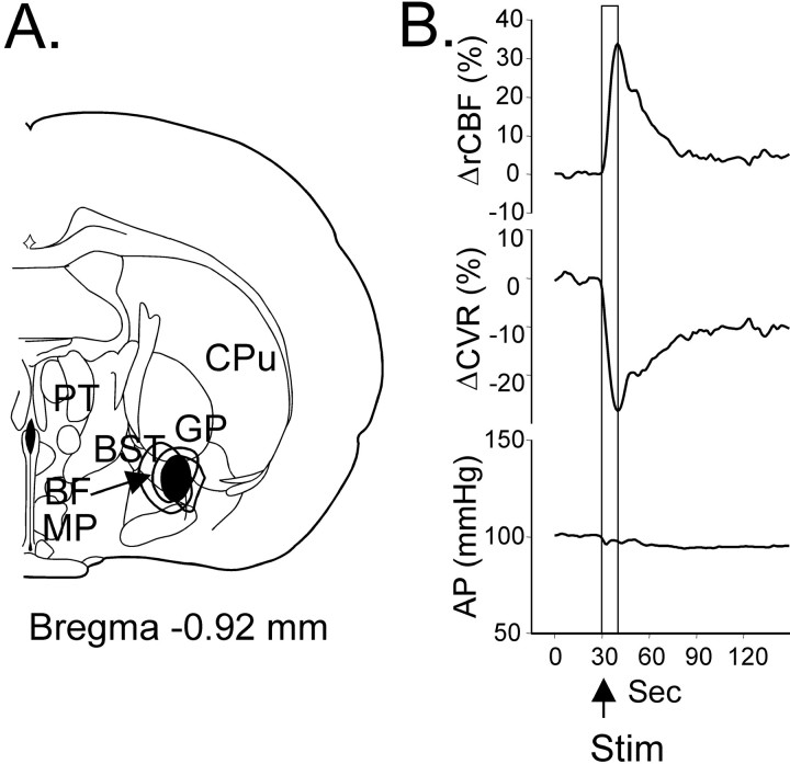
Fig. 8.
Localization of the lesion sites in the BF (A) and effect of stimulation of these sites (B) before the lesions on rCBF (top trace), CVR (middle trace), and AP (bottom trace). Contours of all lesions are superimposed, and the area common for all lesions isblackened. Abbreviations are as in Figure 1.

