
Fig. 3.
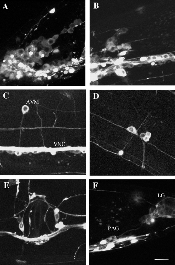
High magnification images showing expression pattern of UNC-104:: GFP in a wild-type transgenic wormejEx52–1. Animals were oriented with their heads on the left side and their dorsal sides facing up. Scale bar, 10 μm.A, Left side of the worm showing the lateral and ventral ganglion; B, retrovesicular ganglion; C, AVM neuron and ventral nerve cord (VNC);D, left posterior lateral ganglion; E, vulva; F, preanal ganglion (PAG) and lumbar ganglion (LG).