
Fig. 3.
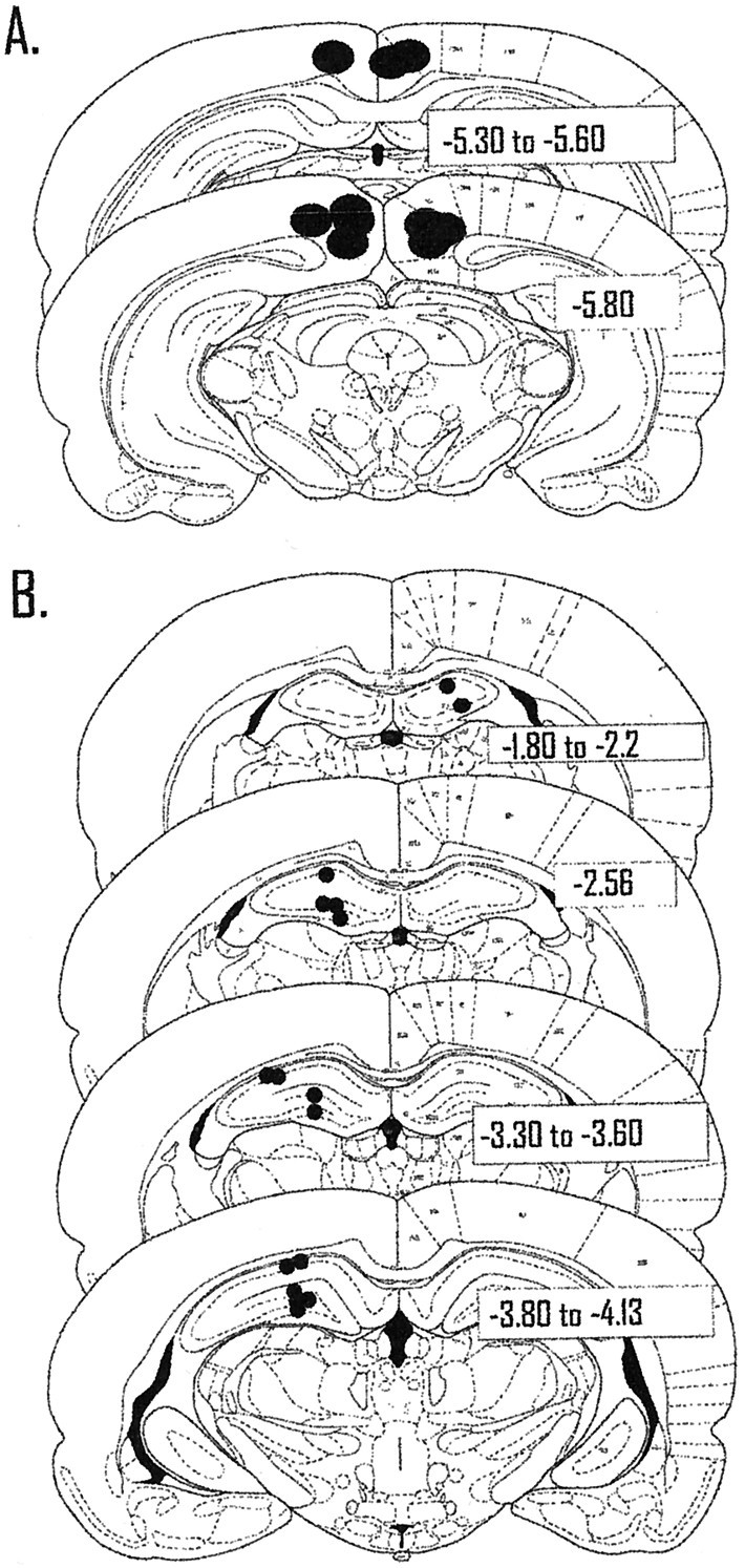
Location of the tips and recording sites of the guide cannulas. A, Each filled circlecorresponds to a single guide cannula. Previous work and ink injections have shown that the spread of tetracaine is just slightly more than a 1 mm circumference around the injection site. Therefore, retrosplenial granular and agranular areas, cingulum bundle, and Oc2MM of posterior parietal cortex were likely affected by injections of tetracaine. B, Each filled circlecorresponds to two to eight cells recorded in that location. For electrode tracks that passed through the same area in different animals, a single filled circle is used to signify the recording site of multiple cells. The majority of cells (n = 43) were recorded from CA3 in the left hemisphere; a smaller number of cells (n = 15) were also recorded in CA1.