
Fig. 1.
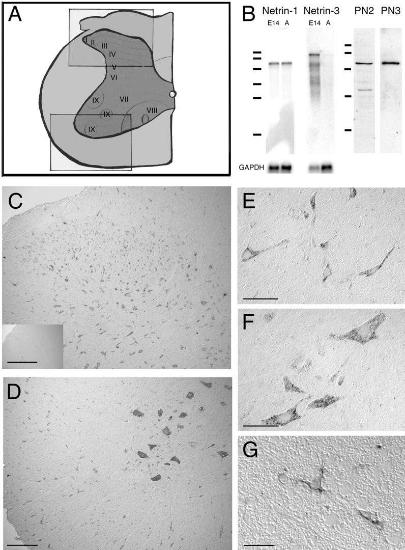
Distribution of netrin-1-expressing cells in adult rat spinal cord. A, Illustration of a hemisection of an adult rat spinal cord [adapted from Paxinos (1995)] is shown. Theboxes correspond to the regions displayed in the micrographs of dorsal spinal cord (C, top box in A) and ventral spinal cord (D, bottom box inA). B, Northern blot analysis of E14 rat brain and adult rat spinal cord poly(A+) RNA (2 μg of RNA) identified a single ∼6 kb mRNA transcript encoding netrin-1, and for netrin-3 identified a major transcript at ∼9 kb and several minor transcripts only in the E14 brain. Netrin-3 is not expressed at detectable levels in the adult spinal cord. RNA size standards correspond to 9.49, 7.46, 4.40, 2.37, 1.35, and 0.24 kb (Bio-Rad, Hercules, CA). Western blot analyses of protein present in a high-salt extract of the membrane fraction of adult rat spinal cord homogenate using antibodies PN2 or PN3 are shown. Both antibodies reveal an ∼75 kDa immunoreactive band, consistent with the molecular weight of netrin-1. The additional minor lower-molecular weight immunoreactive bands may be proteolytic fragments of full-length netrin protein. Protein size standards correspond to 116, 97.4, 66.2, 45, and 31 kDa (Bio-Rad). C–G, In situhybridization analysis identified netrin-1-expressing cells in all laminas and the white matter of dorsal (C) and ventral (D) hemisections of C5 spinal cord.E illustrates the morphology of netrin-1-expressing cells in lamina IV of the dorsal horn. Hybridization was detected in the cytoplasm and the proximal portion of neurites. Large neurons in the ventral horn with the morphological characteristics of motoneurons express netrin-1 (D,F). Netrin-1-positive cells were also detected throughout the white matter (C, D). G illustrates the morphology of netrin-1-positive cells located in ventral C5 white matter. Like the neurons in E and F, positive hybridization was detected in the cytoplasm and proximal processes of these glial cells. The small inset in Cillustrates the absence of hybridization using the corresponding sense cRNA probe. C–G, Differential interference contrast (DIC) optics, digoxigenin-labeled probe visualized with a POD-conjugated secondary antibody against digoxigenin and the diaminobenzidine substrate, is shown. Objective magnification:C, D, 10×; E, F, 40×; G, 100×. Scale bars: C, D, 150 μm; E, F, 50 μm; G, 25 μm. GAPDH, Glyceraldehyde-3-phosphate dehydrogenase.