
Fig. 1.
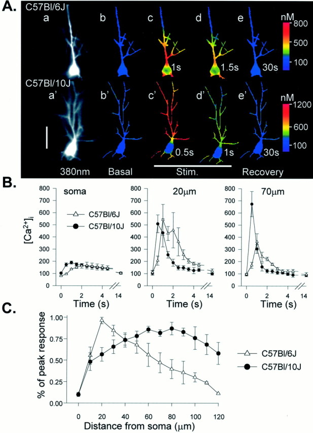
Strain difference in dendritic Ca2+ responses after tetanic stimulation of Schaffer collateral inputs. A, Examples of intracellular Ca2+ transients in Bl/6 (top panels) and Bl/10 (bottom panels) neurons in response to a single tetanus applied to the stratum radiatum (50 Hz, 2 sec). Fluorescence images (a, a′) show bis fura-2 excited at 380 nm. Right panels are ratio images (350/380 nm excitation) showing Ca2+ levels as false color images (calibration at right). Resting levels are shown in b, b′, and subsequent frames were taken at the times indicated after the onset of the tetanus. In the Bl/6 neuron the peak Ca2+ increases during the tetanus (c) were restricted to the proximal apical dendrite and soma before declining during the stimulus (d) and recovering to near-resting levels 30 sec after the stimulus (e). In contrast, the same stimulus produced substantially larger Ca2+ transients in the distal dendrites of the Bl/10 neuron, and peak responses throughout the Bl/10 neuron occurred sooner than transients in the Bl/6 cell (c′) before recovery to resting levels (d′, e′). Scale bar, 50 μm. B, Group data showing the time course of Ca2+ concentrations at three locations in response to 50 Hz, 2 sec tetani. Peak concentrations are similar in soma and proximal dendritic regions of both groups (soma and 20 μm) but substantially larger in Bl/10 neurons at more distal sites (70 μm). Dendritic Ca2+ signals also peak earlier after the onset of the tetanus in the Bl/10 strain (20 μm and70 μm; n = 6 Bl/10 neurons andn = 5 Bl/6 neurons). C, Responses normalized to peak responses in each cell, illustrating that peak Ca2+ responses in Bl/6 neurons were observed close to the soma in the proximal apical dendrite, but peak responses in Bl/10 cells were observed much further from the soma (n = 6 and 5 for Bl/10 and Bl/6, respectively).