
Fig. 3.
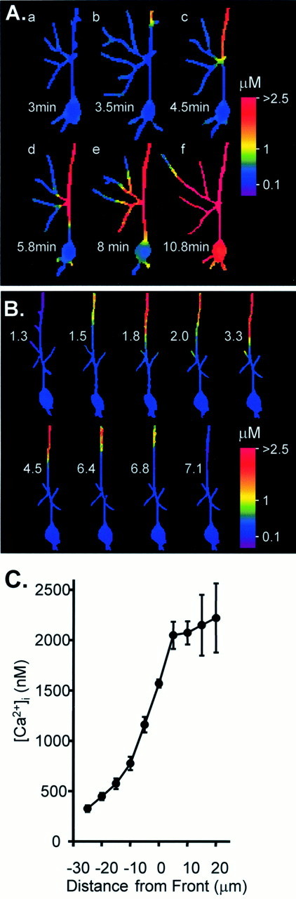
Secondary Ca2+ responses in Bl/10 dendrites after repetitive bolus KA exposure.A, Ca2+ images at the times indicated after washout of the third of three KA bolus applications. Dendrites within the field of view had recovered to resting levels 3 min after KA washout (a). With the passage of time a secondary response invaded the dendrite within the field of view (b, top). The response then propagated along the apical dendrite and invaded secondary dendrites as they were encountered (c, d) before arriving at (e) and involving the cell soma (f). B, A secondary Ca2+ response was generated by six bolus KA applications. The response originated in distal dendrites, progressed toward the soma, and then receded until the neuron recovered to prestimulus Ca2+ levels. The numbersin each panel indicate the time (in minutes) after the initial peak Ca2+ response in the proximal apical dendrite during the final KA exposure. C, Mean data illustrating the steep Ca2+ concentration across the front of propagating secondary responses. Data were derived from regions 50–100 μm from the cell soma after secondary responses were initiated in the distal dendrites (n = 9 Bl/10 neurons).