
Fig. 4.
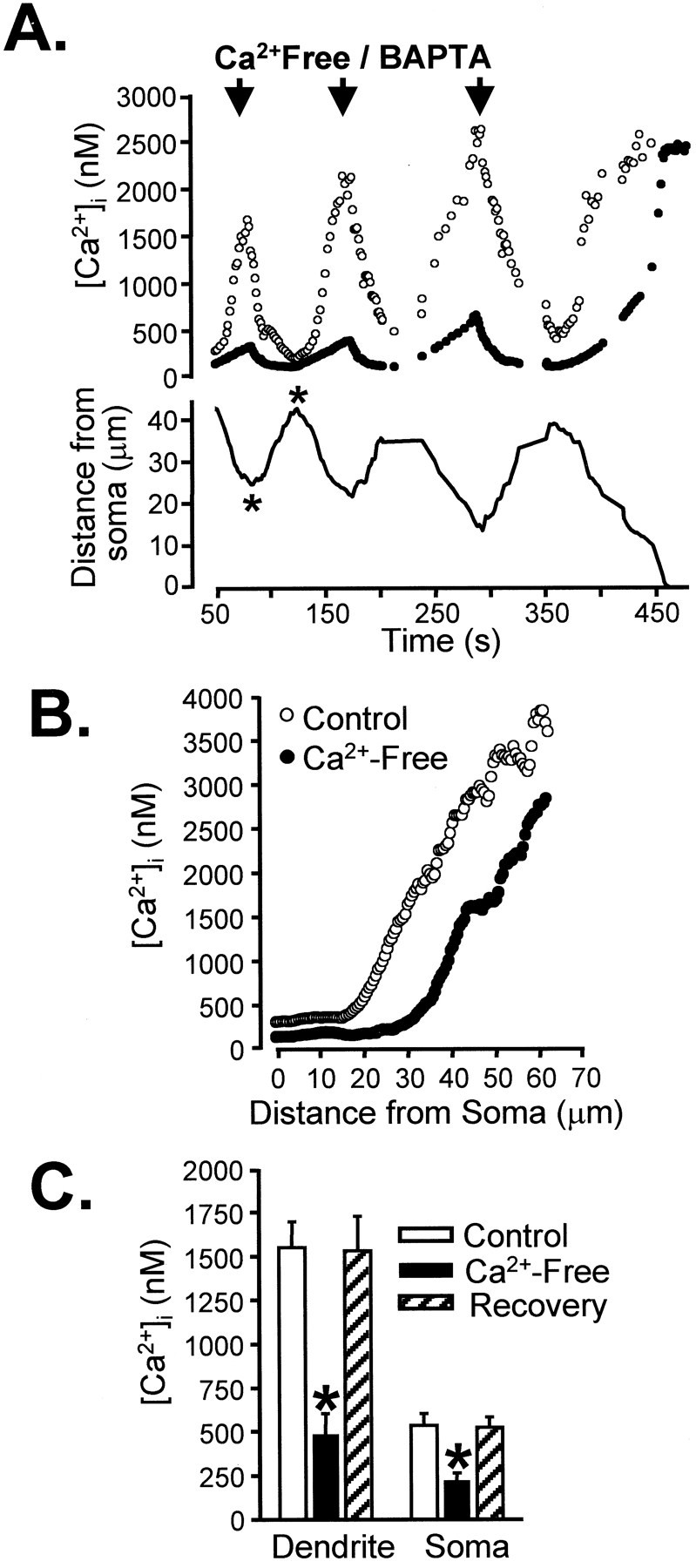
Rapid localized reduction of extracellular [Ca2+] quenches secondary intracellular [Ca2+] increases in Bl/10 neurons.A, Shown are the effects of three successive pressure applications of BAPTA/zero Ca2+ solution (arrows) applied during the progression of a secondary response toward the soma. Dendrite Ca2+ measured ∼30 μm from the soma (open circles) dropped sharply and then recovered after each BAPTA/zero Ca2+application. As a consequence of the close proximity of the dendrite secondary response, soma Ca2+ levels also were elevated significantly (filled circles) and also were reduced during BAPTA/zero Ca2+ application. The position of the leading edge of the secondary response is indicated by the solid line and shows that, after the three zero-Ca2+ applications, the secondary response was allowed to proceed into the cell soma. B, Profile of Ca2+ concentrations as a function of distance from the soma at the times marked by the asterisks inA. C, Mean data from a group of five neurons showing mean Ca2+ concentrations in soma and dendrite (30–50 μm from soma) before, during, and 2 min after a brief (3 sec) BAPTA/zero Ca2+ exposure (*p < 0.005).