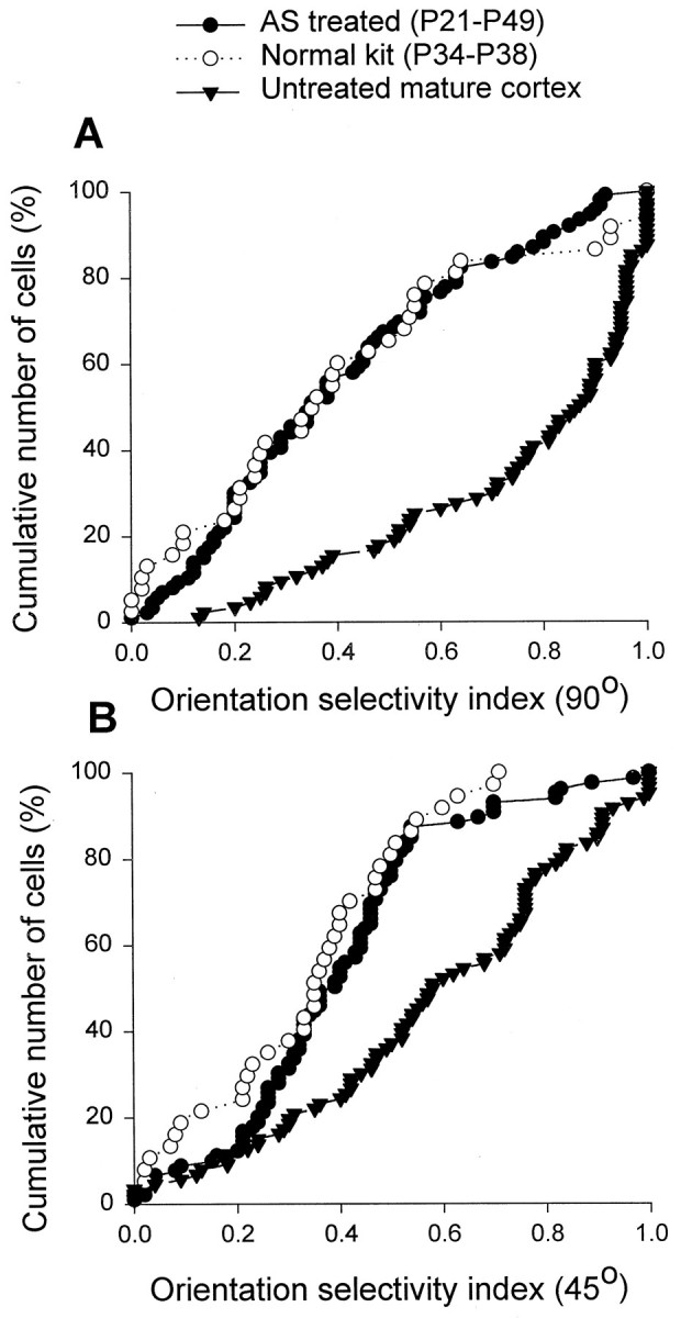
Fig. 3.

Antisense ODN treatment prevented the developmental changes in the orientation selectivity indices of cortical cells. The cumulative percentage of cells was plotted as a function of the orientation selectivity index at 90° (A) and 45° (B) for three groups of animals: (1) antisense ODN-treated and studied at ∼P49, (2) untreated kit studied around the time of eye opening, and (3) untreated mature animal studied around the same age as the antisense ODN-treated animals. The orientation selectivity indices for the cells studied in the untreated animals increased markedly from the time of eye opening until maturity at P49 (compare the open symbols andfilled triangles). In contrast, the antisense ODN-treated animals studied at P49 had orientation selectivity indices that were similar to those found in kits but markedly lower than those found in mature untreated ferrets. The distributions for antisense ODN-treated animals and untreated animals are different statistically (Wilcoxon–Mann–Whitney U test; p< 0.01). In contrast, the distributions for antisense ODN-treated animals and the untreated kittens are indistinguishable (p > 0.05).
