
Fig. 2.
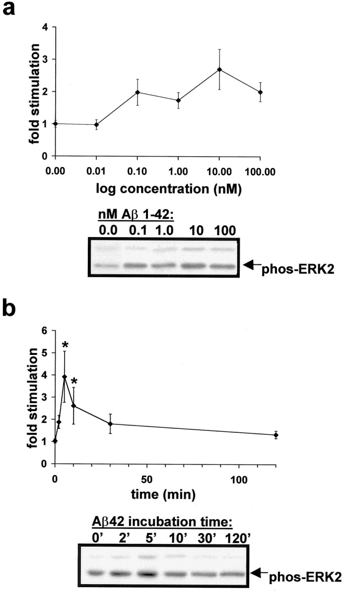
Time course and concentration dependence of Aβ42 activation of ERK2 MAPK in cultured rat hippocampal slices.a, Aβ42 activates ERK MAPK in the picomolar to nanomolar range. Data points are as follows: 0 nm(1.00 ± 0.15), 0.01 (0.98 ± 0.16), 0.1 (1.98 ± 0.48), 1.0 (1.74 ± 0.25), 10 (2.70 ± 0.62), and 100 (2.01 ± 0.30) nm Aβ42 for 5 min. Representative immunoblot results are depicted below the response curve.b, The 100 nm Aβ42 rapidly activates ERK2 MAPK in rat hippocampal slice cultures. Peak response occurs at 5 min Aβ42 and returns to baseline within 2 hr. Data points are as follows: 2 (1.88 ± 0.30), 5 (3.92 ± 1.15), 10 (2.61 ± 0.83), 30 (1.81 ± 0.44), and 120 (1.34 ± 0.17) min. *Significant difference from basal level (1.06 ± 0.12); p< 0.05, Student's t test with Welch's correction because variances differ significantly according to Bartlett's test. Representative immunoblot results are depicted below the response curve.