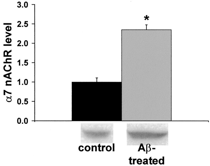
Fig. 7.
Chronic exposure to Aβ42 leads to increased α7 nAChR protein in hippocampal slice cultures. Cultured rat hippocampal slices were exposed to 100 pm Aβ42 for 144 hr, and α7 nAChR protein was quantified by immunoblot. The data that are expressed are normalized to the α7 nAChR protein level in cultures that were left untreated. Representative immunoblot results are shownbelow the histogram. *Significant difference from control level (p < 0.0001) by Student'st test. Basal, 1.00 ± 0.11; Aβ42-treated, 2.35 ± 0.13.

