
Fig. 6.
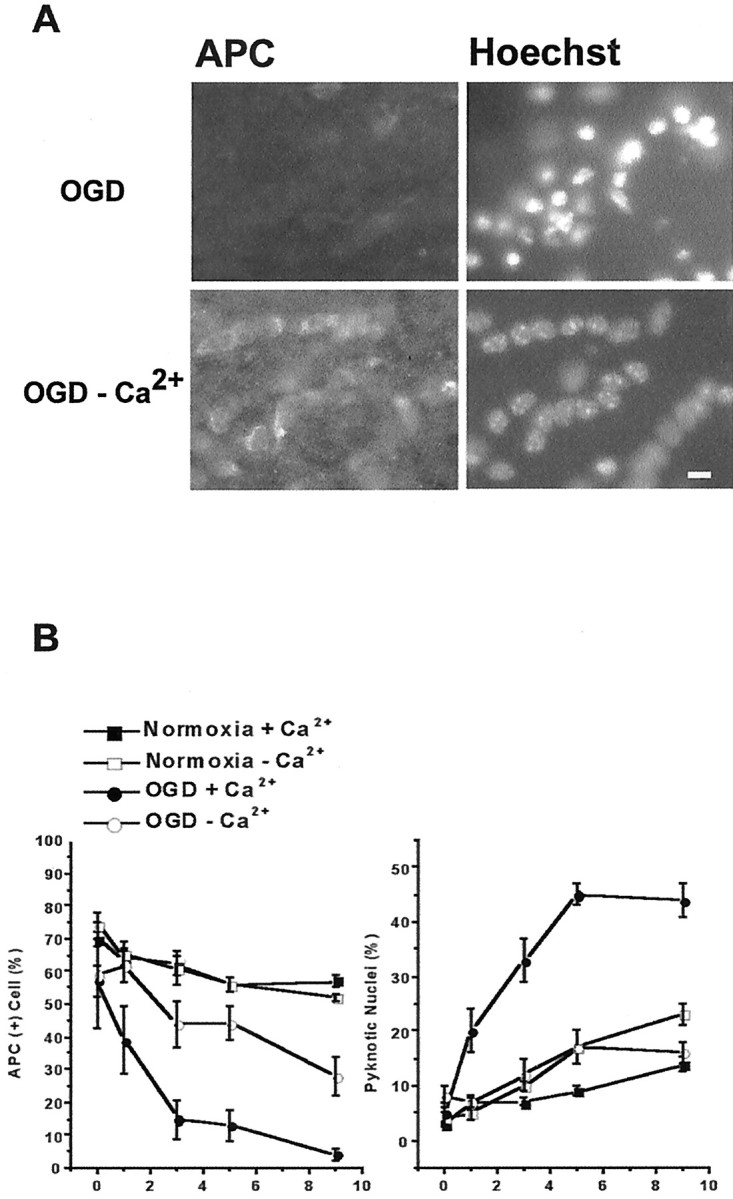
OGD-induced oligodendrocyte death depends on extracellular Ca2+. Slices are exposed to 30 min OGD in normal aCSF (containing 2.0 mm Ca2+) or aCSF with no Ca2+ including 200 μmEGTA. Perfusion conditions are maintained for 30 min before and during and 30 min after OGD. A, APC immunofluorescence of slices fixed 9 hr after OGD. Exposing slices to OGD in Ca2+-free media reduces oligodendrocyte death (left) and pyknotic nuclei formation (right) compared with OGD with normal Ca2+. Scale bar, 10 μm. B, Oligodendrocyte cell counts in slices from normoxia (filled squares), Ca2+-free normoxia (open squares), OGD (filledcircles), and Ca2+-free OGD (open circles) are summarized in the plot on the left. Ca2+-free aCSF in normoxic conditions does not alter oligodendrocyte numbers. OGD in Ca2+-free aCSF remarkably reduces OGD-induced oligodendrocyte death. B,Right, Pyknotic nuclei number in slices exposed to Ca2+-free OGD is significantly less than in slices exposed to OGD. Values represent mean ± SEM.