
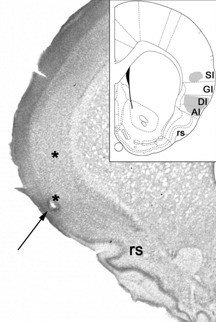
Fig. 1.
Localization of electrode bundles in rat SI and GC. The arrow points to the hole just above the rhinal sulcus, where the microwire tips rested at the time of perfusion. Theasterisks above this site mark the approximate position of the electrode tips when GC and oral SI recordings were made.Inset, The shaded areas in this schematic, adapted from Paxinos and Watson (1997), demarcate the limits of the regions from which recordings were made. GI, Granular insular cortex; DI, dysgranular insular cortex;AI, agranular insular cortex; SI, oral somatosensory cortex; rs, rhinal sulcus.