
Fig. 7.
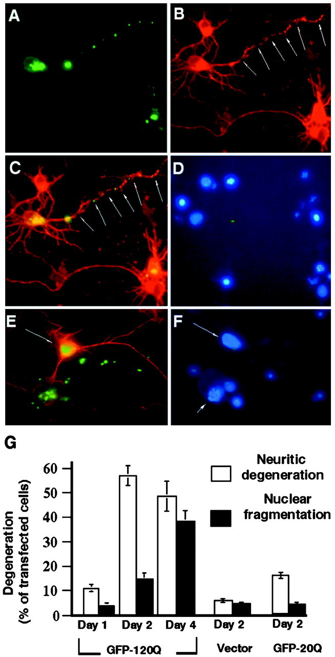
Neuritic degeneration of huntingtin-transfected striatal neurons before nuclear fragmentation. A–D, Cells were transfected with GFP-120Q for 24 hr and then stained with antibodies to huntingtin (A) and tubulin (B). In the merged image(C), neuritic aggregates are associated with the fragmentation of neurites that display discontinuous tubulin labeling.D, Nuclear staining did not show any DNA fragmentation in the neuron that has degenerated neurites. Small nuclei (no arrows) of glial cells are also shown. E, F, Amerged image of striatal neurons transfected with GFP-120Q for 48 hr. The cells were stained with antibodies against huntingtin (green) and tubulin (red). Note that a cell containing intranuclear huntingtin has intact nuclear staining (arrow) and neuritic tubulin labeling. A transfected cell showing DNA fragmentation (arrowhead), however, displays neuritic aggregates and has lost neuritic tubulin staining. G, Quantitative measurement of GFP-120Q-, GFP-20Q-, or GFP vector-transfected cells that displayed neuritic degeneration and nuclear degeneration. The data were obtained by counting transfected cells in two to three independent transfections.