
Fig. 8.
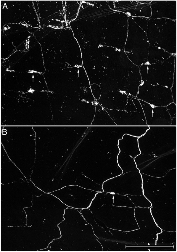
There was a substantial loss of IGLEs in the duodenum of NT-4-deficient mice. Low-magnification darkfield photomicrograph of IGLEs labeled with WGA-HRP (some IGLEs are identified by arrows) in a wild-type (A) and an NT-4 mutant (B) mouse. Each field is located in the same region of the duodenum, a region that normally has a high density of IGLEs. Only one labeled IGLE is present in this region of a mutant duodenum (B). Scale bar, 0.5 mm.