
Fig. 2.
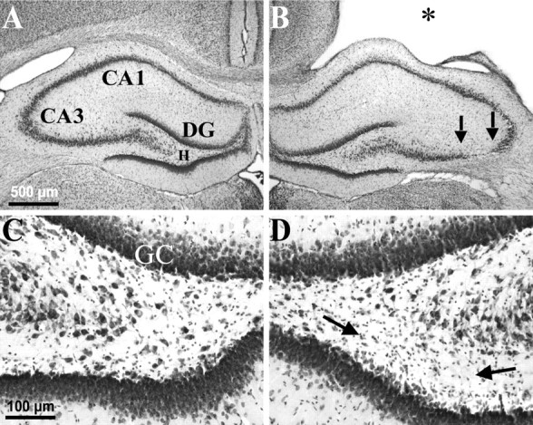
Weight-drop TBI-induced selective, gross cell loss in the ipsilateral hippocampus. A, A cresyl violet-stained coronal section 3 weeks after TBI shows the hippocampus contralateral to TBI with no evidence of macroscopic cell loss.B, From the same brain as A, showing the hippocampus ipsilateral to TBI. Note the TBI-induced cortical cavity (∗). Gross CA3 cell loss is delineated by arrows. There was no obvious cell loss in the CA1 or dentate granule cell layers. C, A higher magnification from Ashows the contralateral hilus with no evidence of gross cell loss after TBI. D, A higher magnification from Bshows the ipsilateral hilus where a subtle loss of large cells can be detected, especially ventrally (between arrows) in this example, as compared with the contralateral side in C.DG, Dentate gyrus; GC, granule cell layer; H, hilus of the dentate gyrus.