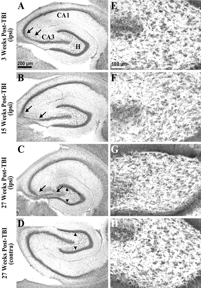
Fig. 3.

Gross cell loss in the ipsilateral hippocampal CA3 and hilus progressed in temporal regions during the weeks after TBI. A, A cresyl violet-stained horizontal section of the temporal hippocampus from ∼4.5 mm deep with respect to bregma prepared 3 weeks after TBI shows gross cell loss in the ipsilateral CA3 (between arrows). Otherpanels on the left show similarly prepared sections. B, The ipsilateral hippocampus 15 weeks after TBI showed a wider region of gross cell loss in CA3 (between arrows) compared with A.C, The ipsilateral hippocampus 27 weeks after TBI showed a progression of cell loss across the entire CA3. Note atrophy of the ipsilateral hilus, indicated by reduced distance between supragranule and infragranule cell layers (arrowheads) compared withD. D, The contralateral hippocampus 27 weeks after TBI (from the same brain section as C) showed no evidence of gross cell loss.E–H show higher magnifications of hilus from A–D. E, At 3 weeks after TBI, cell loss was subtle in the ipsilateral hilus in this temporal location. F, At 15 weeks after TBI, cell loss remained subtle in the temporal ipsilateral hilus. G, At 27 weeks after TBI, cell loss and atrophy of the temporal ipsilateral hilus was clearly detectable, although some large hilar neurons remain.H, There was no evidence of gross cell loss in the contralateral hilus 27 weeks after TBI at this temporal location. Scale bars in A and E apply toA–D and E–H, respectively.
