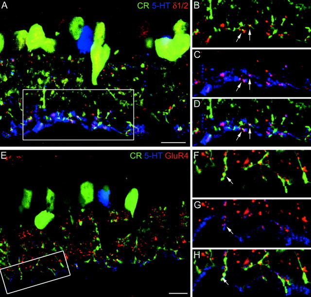
Fig. 7.
A, Confocal fluorescence micrograph of a vertical section through the inner part of a rabbit retina that was triple-labeled for calretinin (CR;green), serotonin (5-HT;blue), and the δ1/2 subunit (red). The inner IPL (frame) is shown at higher magnification in B–D. B, The green AII cell dendrites and the redδ1/2 hot spots are not in register. Only two dots(arrows) coincide with green dendrites.C, The blue, 5-HT-labeled dendrites of AI amacrine cells coincide with the red δ1/2 hot spots, and as a consequence they appear purple.D, All three markers superimposed. The two dots (arrows) appear white.E, Confocal fluorescence micrograph of a vertical section through the inner part of a rabbit retina that was triple-labeled for calretinin (CR,green), serotonin (5-HT,blue), and GluR4 subunit (red). The inner IPL (frame) is shown at higher magnification inF–H. F, Thegreen AII cell dendrites and the redGluR4 puncta are often in register. G, Theblue, 5-HT-labeled dendrites of AI cells are not in register with the GluR4 puncta. H, All three markers superimposed. The dot indicated by thearrow colocalizes both 5-HT and CR. Scale bars:A, E, 10 μm.

