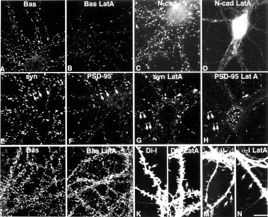
Fig. 8.

In 12-d-old neurons (A–H), Bassoon-labeled clusters (A, Bas) are diminished in size and number after latrunculin A treatment (B, Bas LatA), but are still readily detected. N-cadherin-labeled clusters (C, N-cad) are lost after latrunculin A treatment (D, N-cad LatA), but a few tiny puncta remain (D). In control neurons, PSD-95 labeling (F) codistributes with a subpopulation of synaptophysin boutons (E,syn, arrows). After latrunculin A treatment (G, H), many synaptic PSD-95 puncta are retained (G, H,arrows), but many PSD-95 puncta are nonsynaptic, which is particularly evident in the cell soma (H). In 20-d-old neurons, latrunculin A treatment does not affect the distribution of Bassoon (J) compared with untreated control neurons (I). Di-I labeling reveals both spine-like and filopodia-like protrusions in control neurons (K). After latrunculin A treatment, nearly all protrusions appear filopodia-like (L). α-internexin (α-I) labeling is concentrated in axons, but also appears in dendrites where it can be seen to invade protrusions (M). After latrunculin A treatment, α-internexin labeling remains at the base of dendritic protrusions (N). Scale bar:A–J, 20.2 μm;K–N, 11.3 μm.
