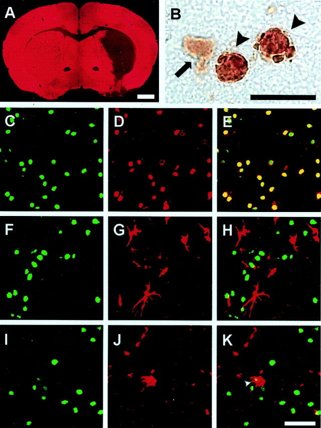
Fig. 1.

Selective neuronal death in the mouse striatum 72 hr after an episode of 30 min MCAo and reperfusion. The low-power view of MAP-2 immunostaining indicates striatal lesion and cortical sparing (A). At higher magnification (B), TUNEL-positive cells (arrowheads) show in DAB staining condensed nuclei with clumped chromatin. Cells showing weak diffuse DAB-positive cytoplasmic staining are not considered TUNEL positive (arrow). Sections were double stained for TUNEL (C,F, I) and cell type-specific markers NeuN (D), GFAP (G), or MAC-1 (J) and examined in a confocal microscope. Cell-specific labeling was visualized using antibodies conjugated with fluorescein (C, F,I: green) or Texas Red (D,G, J: red). No double labeling was detected for TUNEL and the astrocytic marker, GFAP (H). Moreover, there was no double labeling for TUNEL and the microglial marker, MAC-1 (K), with the exception of some cells that most likely represent engulfed nuclei of dead neurons (K,arrowhead). By contrast, most of the TUNEL-positive cells were also immunoreactive for the neuronal marker NeuN (E), indicating neuronal origin of the TUNEL-positive cells. Scale bars: A, 1 mm;B, 10 μm; C–K, 30 μm.
