Fig. 2.

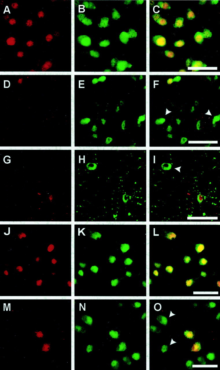
Expression of p16INK4a and p27Kip1, endogenous inhibitors of cyclin-dependent kinases, after 30 min MCAo and reperfusion in the mouse striatum (A–I, p16INK4a) and after OGD in rat primary cortical neurons (J–O, p27Kip1). Immunoreactivity for p16INK4a/p27Kip1was visualized with Texas Red (A, D,G, J, M:red), and neuronal marker NeuN was visualized with Alexa 488 (B, E, H,K, N: green), with colocalization resulting in a yellow color (C, F, I,L, O). Strong nuclear expression of p16INK4a was seen in all neurons in the normal (non-ischemic) striatum as shown by double labeling with p16INK4a and NeuN (A–C). p16INK4aimmunoreactivity was lost in ischemic striatal neurons at 9 hr after MCAo/reperfusion as shown by the appearance of NeuN-positive p16-negative cells (D–F,arrowheads). Double labeling of p16INK4a with the ischemia-sensitive neuronal marker MAP-2 demonstrated that the loss of p16INK4aexpression occurred in cytoarchitectonically intact neurons at 9 hr (G–I; arrowhead inI). Strong nuclear p27Kip1immunoreactivity was detected in all neurons in primary neuronal culture (J–L). Two hours after OGD the majority of neurons downregulated p27 Kip1, as indicated byarrowheads (M–O). Scale bars, 30 μm.
