Fig. 4.

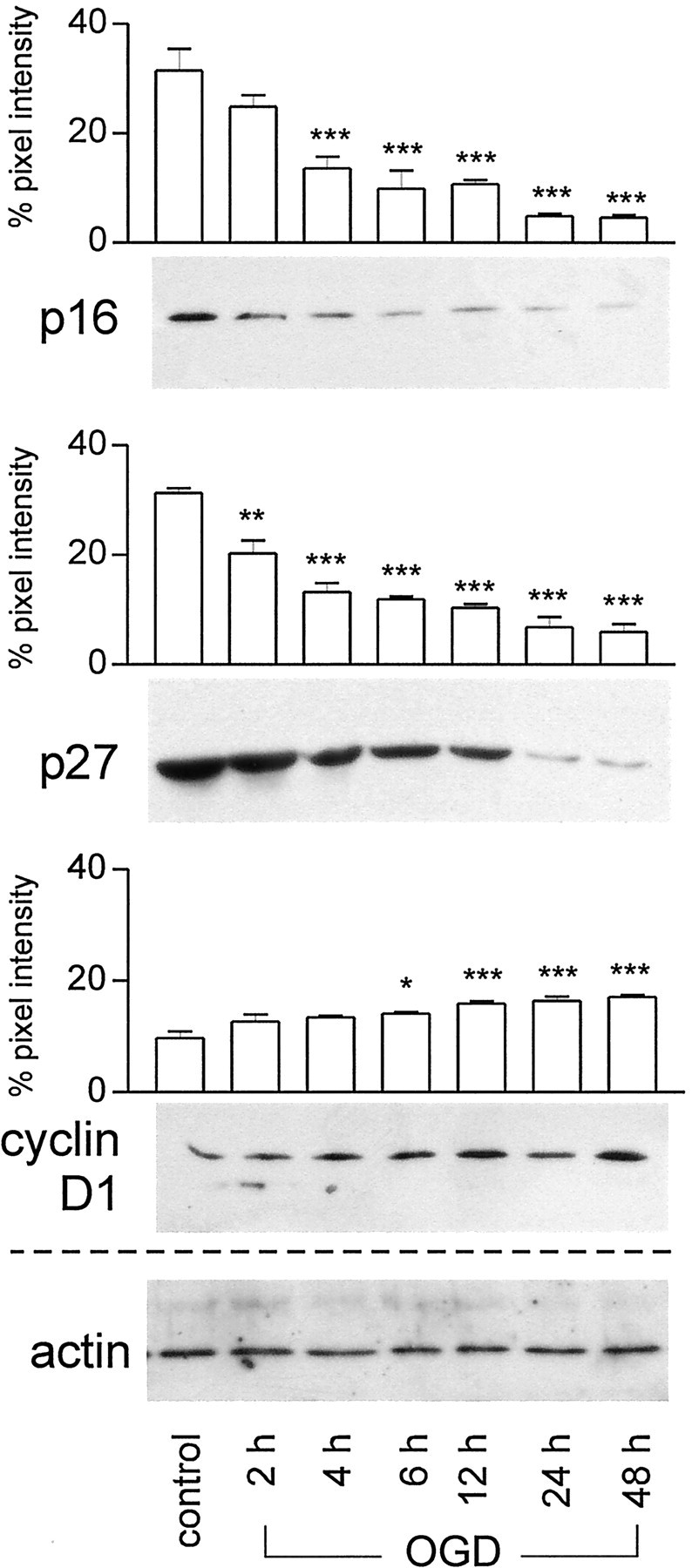
Immunoblots showing time-dependent changes of cell cycle-related proteins in primary cortical neurons after 90 min oxygen–glucose deprivation. Cell lysates (20 μg) were subjected to SDS-PAGE, and membranes were probed with antibodies against p16INK4a, p27Kip1, and cyclin D1 (0.2–1.0 μg/ml). Actin served as internal control. The experiment was repeated three times; a representative experiment is shown. For semiquantitative analysis, the intensity of each band was quantitated from scanned images of nonsaturated immunoblot films using Scion Image (Scion Corporation, Frederick, MD). The pixel intensity of the bands obtained in each experiment was summed and set as 100%, and the individual band was calculated as percentage of total signals. The graphs show a significant downregulation of p27Kip1starting at 2 hr and of p16INK4a at 4 hr after OGD compared with controls. Cyclin D1 levels were significantly upregulated at 6 hr and further increased at 48 hr. Mean value ± SEM. *p < 0.05; **p < 0.01; ***p < 0.001.
