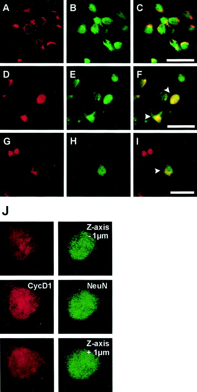
Fig. 5.

Cyclin D1 expression in primary cortical neurons in culture after OGD (A–F) and in striatal neurons of mice after MCAo/reperfusion (G–I). Immunoreactivity for cyclin D1 was visualized with Texas Red (A,D, G; red), and neuronal marker NeuN was visualized with Alexa 488 (B,E, H; green). Double labeling for cyclin D1 and NeuN demonstrates that cyclin D1 is expressed exclusively in the cytoplasm of cultured neurons under control conditions (A–C). Two hours after OGD cyclin D1 is strongly upregulated, and its immunoreactivity translocates to the nucleus (D–F,arrowhead). Confocal Z-series images (step = 1 μm) confirm the nuclear expression of cyclin D1 in OGD-treated neurons (J). Cyclin D1 is not expressed in normal striatal neurons (data not shown). Forty-eight hours after MCAo/reperfusion, nuclear cyclin D1 immunoreactivity is detected in neurons in the ischemic striatum (G–I,arrowhead). Scale bars:A–I, 30 μm.
