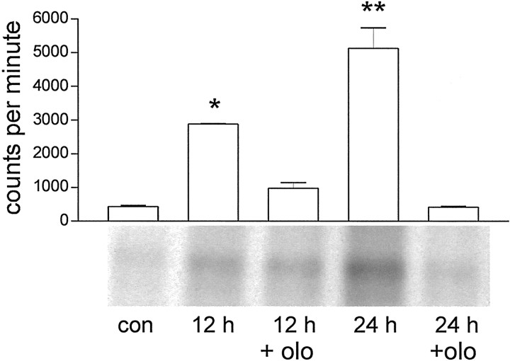
Fig. 6.
Cdk2 activity increases in cortical neurons in culture after 90 min OGD. Cell lysates were immunoprecipitated with anti-Cdk2 antibody, and the resultant complexes were allowed to incubate with [γ-32P]ATP and histone HIIIS as substrate. In each lane, 30 μg protein was separated. There was a clear increase in kinase activity at 12 and 24 hr after OGD as compared with control. This kinase activation at 12 and 24 hr was completely abolished when the cells were pretreated with 10 μmolomoucine (olo) 1 hr before and during OGD. To estimate Cdk2 activity, the amount of incorporated radioactive label was quantified using a phosphorimager and TINA software. The values of three independent experiments are graphically represented as mean value ± SEM. *p < 0.01; **p < 0.001 versus control.

