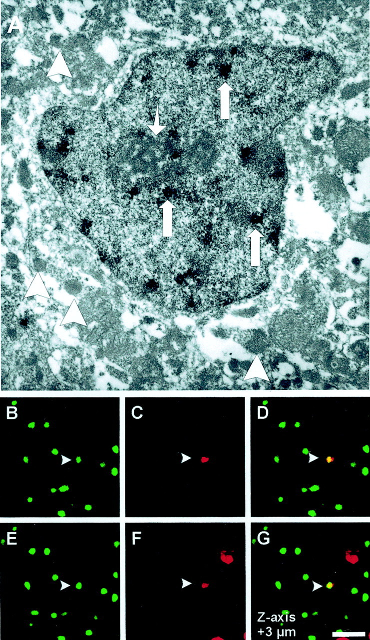
Fig. 8.

TUNEL/BrdU double labeling. BrdU was administered via subcutaneous osmotic mini-pumps in 129/SV mice (1 mg · hr−1 · kg−1 body weight). Mice were subjected to 30 min of MCAo and 72 hr reperfusion.A, For electron microscopy studies, Vibratome sections of ischemic striatum were immunostained for BrdU using DAB as a chromagen. Electron micrograph shows a cell with numerous electron-dense osmiophilic granules (large arrows) within the nucleus corresponding to the BrdU labeling. The cell has a central nucleus with a prominent nucleolus (small arrow) and multiple vesicles (arrowheads) in its cytoplasm and lacks glial filaments, indicating that it is of neuronal origin. Original magnification: 11,000×. TUNEL/BrdU double labeling was performed on fresh-frozen cryosections (10 μm) using ApopTag Kit and rat monoclonal anti-BrdU antibody. TUNEL was visualized with fluorescein (B, E: green), and Texas Red was used for BrdU immunoreactivity (C,F: red). Of all TUNEL-positive cells, 0.957 ± 0.172% stain positive for the S-phase marker BrdU (n = 4 animals). Z-series of confocal images through the nucleus (1 μm steps) confirms the costaining for both markers (D, G: yellow). Scale bar, 30 μm.
