
Fig. 3.
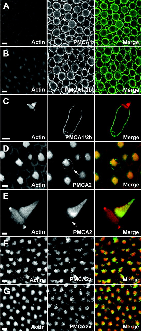
Localization of bullfrog sacculus PMCA isozymes by immunofluorescence. Left columns, Actin (FITC-phalloidin); middle columns, PMCA; right columns, combined actin (red) and PMCA (green). A, F1N labeling for PMCA1. Shown is a cross section through a bullfrog sacculus; plasma membranes of hair cells and supporting cells (arrow) are labeled. B, Fb labeling for PMCA1b and PMCA2b. Shown is a cross section through bullfrog sacculus hair cells and supporting cells. C, Fb labeling for PMCA1b and PMCA2b. The hair bundle is unlabeled. D, F2N labeling for PMCA2. Shown are apical surfaces of bullfrog sacculus hair cells and supporting cells; there is PMCA2 labeling in stereocilia and in the pericuticular necklace (arrow). E, F2N labeling for PMCA2. Shown is labeling in an isolated hair cell; there is strong labeling near the base of the tallest stereocilia and labeling along the apical surface (arrow). F, F2a labeling for PMCA2a. Shown is a whole-mount view of hair bundles. G, F2v for PMCA2v. Shown is a whole-mount view of hair bundles. Scale bars:A–C, F, G, 10 μm;D, 5 μm; E, 2 μm.