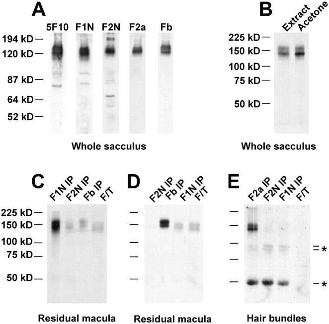
Fig. 5.
Protein immunoblot and immunoprecipitation analysis of bullfrog sacculus PMCA isozymes. A, Immunoblotting of whole bullfrog sacculus with 5F10 and isozyme-selective antibodies. One saccular equivalent was used for each lane. B, 5F10 immunoblot indicating efficiency of acetone precipitation. Extracts from bullfrog sacculi either were added directly to sample buffer (Extract) or were precipitated with acetone (Acetone). Band intensities were similar, indicating that acetone precipitation was efficient. C, 5F10 detection of PMCAs sequentially immunoprecipitated from a residual macula extract (five saccular equivalents) with PMCA-selective antibodies. F/T, Flow through, those proteins that did not bind to any of the precipitating antibodies. D, Same immunoprecipitation and detection conditions as in C, except that the antibody order is changed. E, 5F10 detection of PMCAs sequentially immunoprecipitated from a hair bundle extract (47 saccular equivalents) with PMCA-selective antibodies. Indicated bands (*) were derived from the precipitating antibodies (data not shown).

