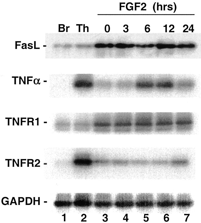
Fig. 4.
Southern blots of RT-PCRs. RT-PCRs were performed using purified mRNA from adult rat brain or thymus (lanes 1, 2) or H19-7 cells (lanes 3–7). H19-7 cells were serum starved in N2 medium at 39°C for 24 hr (lane 3) and then treated with 10 ng/ml FGF2 for 3 hr (lane 4), 6 hr (lane 5), 12 hr (lane 6), or 24 hr (lane 7). The identities of the amplified bands were verified by Southern blotting with the probes described in Materials and Methods. Br, Brain; Th, thymus.

