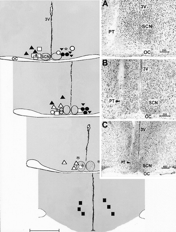
Fig. 1.

Diagrammatic coronal sections showing histologically verified locations of the microdialysis probes tips of different groups included in the study. Coronal planes extend from the anterior SCN (top left section) to the caudal hypothalamus (bottom right section).Symbols represent the ventral extent of the probe implants for the treatment groups as follows: *, p-CPA plus SCN 8-OH-DPAT perfusion; ▾, no p-CPA plus SCN 8-OH-DPAT perfusion; ○, p-CPA plus SCN ACSF vehicle perfusion; ●, p-CPA plus SCN 8-OH-DPAT perfusion in light; ▴, p-CPA plus SCN 5-HT perfusion; ▵, p-CPA plus SCN 8-OH-DPAT plus DR4004 perfusion; ■, p-CPA plus SCN 8OH-DPAT plus ritanserin perfusion; ▪, p-CPA plus 8-OH-DPAT perfusion 2 mm caudal to SCN.OC, Optic chiasma; 3V, third ventricle. Scale bar, 1.0 mm. Insets are photomicrographs of cresyl violet-stained coronal hypothalamic sections showing the location of the probe tip (PT) relative to the SCN in three animals under higher (A, B) and lower (C) magnification. Note the positioning of the microdialysis tip against the lateral margin of the SCN. Values shown above the scale bars are in micrometers.
