Fig. 5.
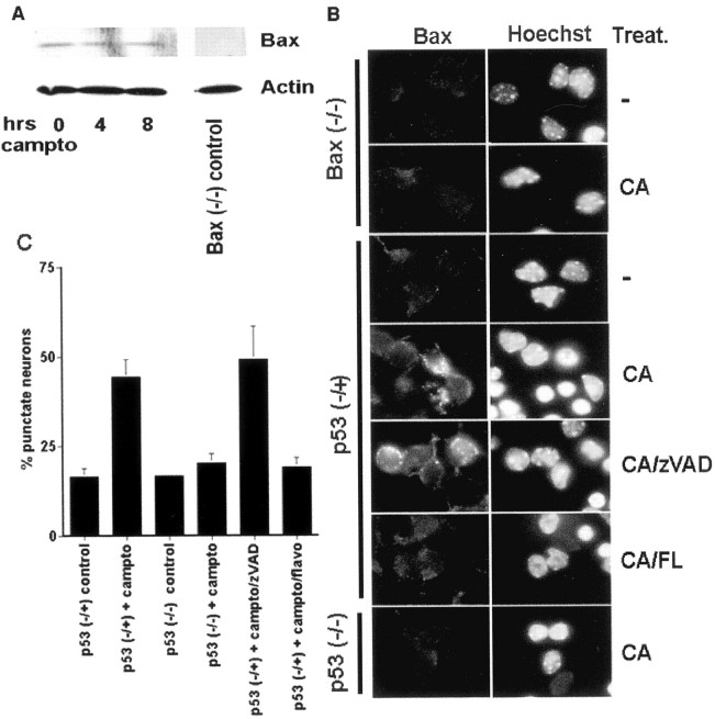
DNA damage-induced Bax translocation is inhibited by either p53 deficiency or CDK inhibition. A, Bax protein levels do not change during the death of cortical neurons evoked by DNA damage. Shown are Western immunoblots of whole-cell lysates of cortical neurons after various periods of treatment with camptothecin (campto; 10 μm). Antibody specificity is demonstrated for Western analysis by using lysates from Bax-deficient neurons. Control for protein loading is indicated by actin expression. B, Immunofluorescence micrograph of Bax translocation in cortical neurons of Bax (−/−), p53 (−/+), or p53 (−/−) backgrounds. Where indicated, the cultures were treated with camptothecin (CA; 10 μm; 10 hr) alone or cotreated with the caspase inhibitor zVAD (100 μm) or the CDK inhibitor flavopiridol (FL; 1 μm). Punctate staining is indicative of Bax translocation from the cytosol and is quantitated in C. Data are the mean ± SEM from three cultures.

